NanoString GeoMX Analysis
GeoMx, developed by NanoString, leverages nCounter® barcoding technology to deliver spatially resolved, digital readouts of up to 96 proteins or RNA targets in a multiplexed assay.
In the GeoMx assay, antibody or RNA probes are coupled to photocleavable oligonucleotide tags. Following hybridization to slide-mounted tissue sections, these tags are released from discrete tissue regions through UV exposure. The released tags are then quantitated using the NanoString nCounter system, generating a spatially resolved digital profile of analyte abundance.
Advantages Captures rare events often missed by bulk experiments through tissue-morphology-guided profiling.
Provides high-plex RNA and protein profiling with precise spatial resolution.
Integration with Imaging GeoMx combines standard immunofluorescence and IHC techniques with digital optical barcoding, enabling highly multiplexed and spatially resolved profiling experiments.
Pre-validated and modular assays offer flexibility across diverse research applications.
Tissue Compatibility Works with both FFPE and fresh frozen samples | Multi-Analyte Detection Supports RNA and protein profiling within the same assay | High Multiplexing Analyze up to 96 targets simultaneously | Single-Cell Resolution Imaging capability down to the cellular level | Quantitative Accuracy Broad 6-log dynamic range for reliable data | Modular Assay Panels Ready-to-use or customizable panels to meet diverse research needs | Flexibility Custom morphological markers or probes can be designed for specific scientific questions | Validated Panels Extensive coverage across immunology, oncology, neurodegeneration, neuroinflammation, and more | Curated Inventory A large selection of pathway- and disease-focused panels, with the option for fully customized probe design.
Strengths & Services
We leverage the NanoString GeoMx Digital Spatial Profiler (DSP) to deliver powerful spatial transcriptomics and proteomics solutions from a single tissue slide. Our platform provides morphological context and integrates seamlessly with current histology and genomics workflows, enabling robust, reproducible, and spatially resolved multi-omic data.
With GeoMx, researchers can precisely select tissue compartments or cell types of interest, quantifying protein and gene expression at high resolution. Significant expression patterns across tissue morphology, gene signatures, and disease biomarkers can be uncovered using advanced statistical analysis. The DSP Data Analysis Suite further streamlines this process, transforming raw data into interactive visualizations and meaningful biological insights within minutes.
:
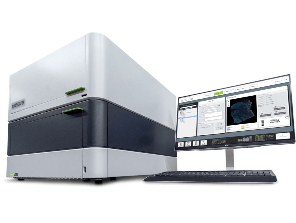
State-of-the-art platforms: GeoMx DSP, nCounter Prep Station, and Analysis System
Rapid turnaround that accelerates discoveries
Pathologist expertise for ROI selection and interpretation
Transparent, collaborative workflow with interactive discussions for study design
30+ years of combined experience in data analysis, interpretation, and scientific support
Flexibility: custom assay design and the ability to quantify any gene/protein of interest, tailored to study needs
Scalability: from small pilot studies to large-scale projects
Comprehensive study reports with actionable insights
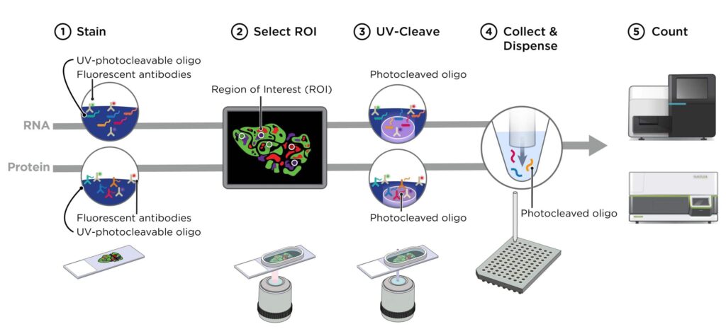
Slides are first prepared and co-stained with fluorescent morphological markers together with oligo-conjugated antibodies for protein assays or oligo-conjugated in situ hybridization (ISH) probes for RNA assays. The samples are then imaged in the GeoMx to visualize morphological markers, enabling region-of-interest (ROI) selection. Each ROI is collected individually by illuminating UV light over the targeted region, which releases photo-cleaved oligos that are subsequently aspirated and counted using the NanoString nCounter system. Finally, the digital counts are mapped back to their original tissue locations, producing a spatially resolved gene and protein profile.
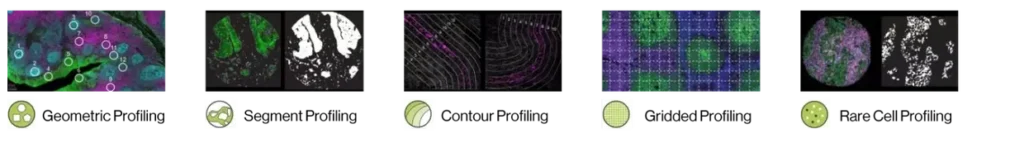
Flexible Profiling Strategies with GeoMx DSP
The GeoMx Digital Spatial Profiler provides multiple strategies for region-of-interest (ROI) selection, giving researchers the flexibility to tailor their analysis to specific biological questions. Geometric profiling enables broad spatial assessment through circular or custom-shaped ROIs, while segment profiling allows separation of distinct tissue compartments such as tumor and stroma. Contour profiling captures expression gradients surrounding defined structures like vasculature or lesions, and gridded profiling generates systematic, unbiased coverage across entire tissue sections. For highly specific investigations, rare cell profiling focuses on unique or low-abundance populations with exceptional precision. Together, these approaches deliver unparalleled flexibility for studying tissue heterogeneity, cellular interactions, and spatial biology.
Have questions? Ask Axela — we’re here to help.
📧 info@axelabio.com